Doppler examination
Doppler examination
Examine blood circulation in mother and child
Doppler sonography (also known as growth ultrasound), a special ultrasound technique, is used to visualise and measure the blood flow in the mother and child.
The blood vessels in the uterus form the maternal supply to the placenta. A significant proportion of serious disorders during pregnancy are due to impaired implantation of the placenta. Successful implantation of the placenta is accompanied by dilation of the blood vessels that supply the intervillous space.
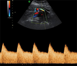
Using the Doppler ultrasound technique, it is possible to analyse the blood flow in the arteries responsible for the uterine blood supply, which ultimately end in the spiral arteries, which in turn release their blood into the intervillous space.
This is where the exchange of blood gases and nutrients with the villous vessels takes place. Doppler blood flow analysis can now be used to determine whether the necessary dilation of the spiral arteries has taken place or not. If this is not the case, in half of the cases considerable problems must be expected in the course of the pregnancy.
By using this non-invasive method, high-risk pregnancies requiring special care can be found with the highest detection rate to date.
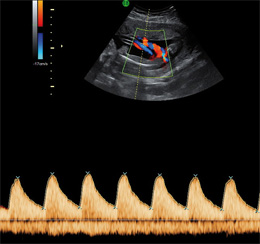
Paediatric examination (fetal vessels):
Doppler examination of foetal blood vessels is the most important method for monitoring an unborn child at risk, e.g. in the event of growth retardation in the womb. Doppler sonography is the only way to carefully monitor foetuses that are too small in order to prevent damage caused by a lack of oxygen.
Today, Doppler sonography is indispensable for monitoring high-risk pregnancies:
- Mothers with high blood pressure
- Mothers with diabetes ("diabetes")
- Twin or triplet pregnancies
- Complicated previous pregnancies
- Various foetal diseases