3-D/4-D sonography

3D ultrasound/4D ultrasound

prenatal diagnostics

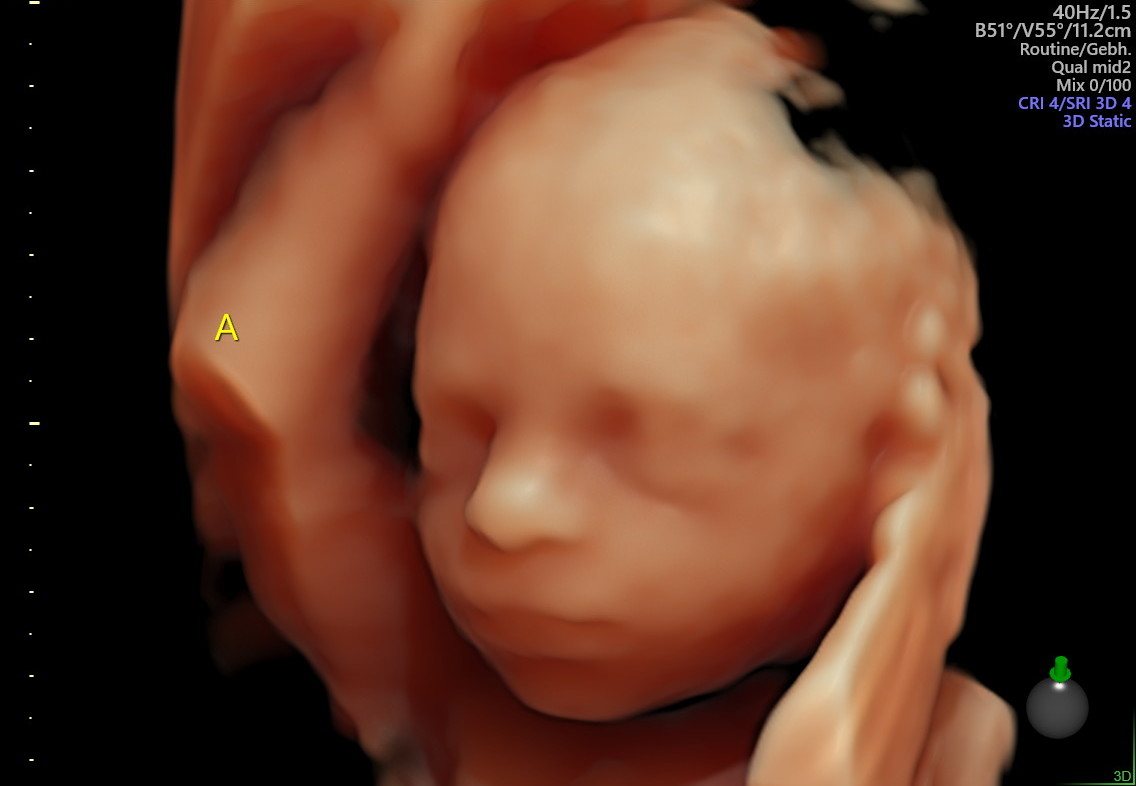
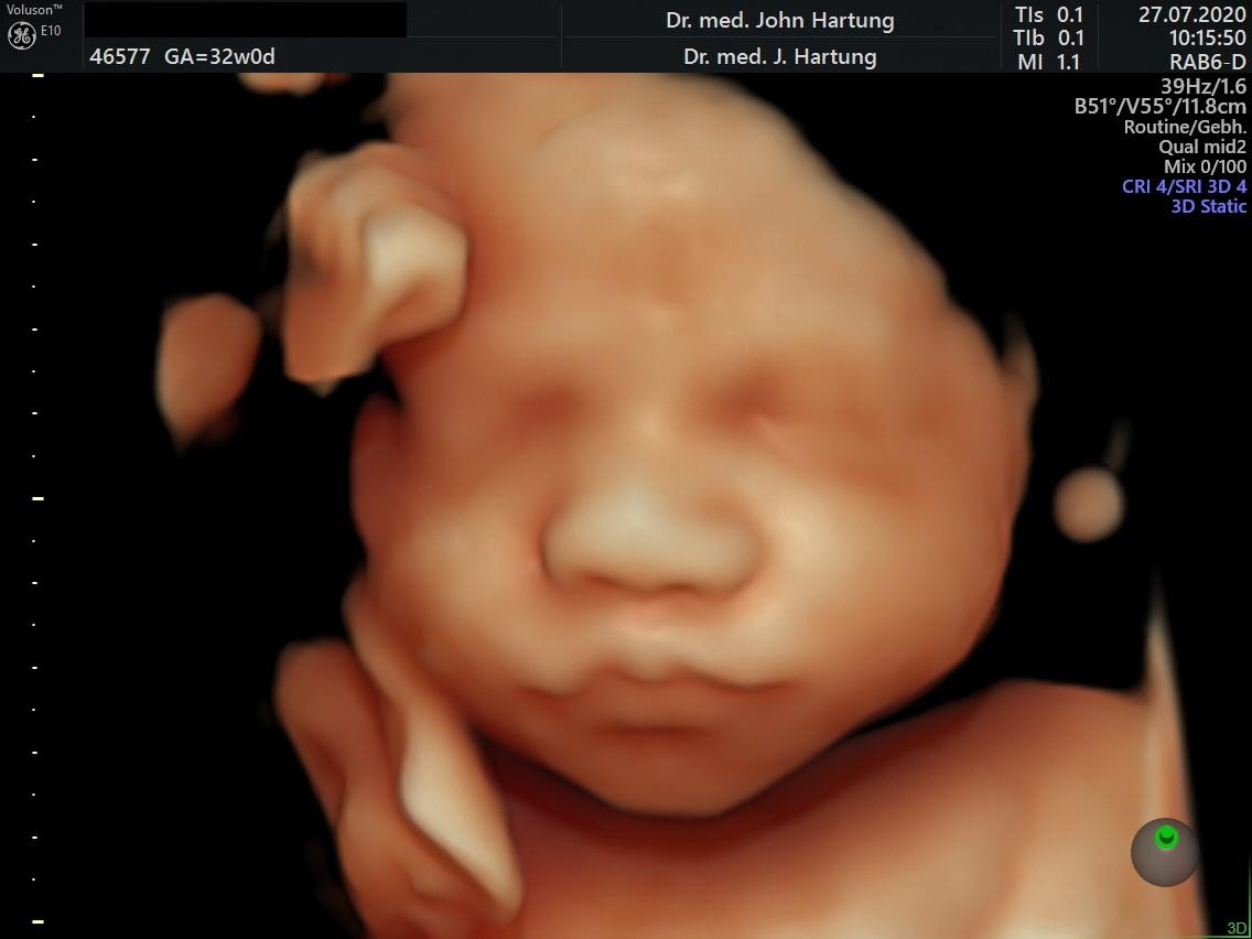
3-D/4-D sonography is the further development of the conventional two-dimensional ultrasound examination in prenatal diagnostics, whereby 3-D stands for a still image and 4-D for moving images. In recent years, the three-dimensional visualisation of foetal surface structures and the multiplanar analysis of organ systems have become an integral part of prenatal ultrasound examinations.

Diseases and malformations can be visualised for patients, but also for paediatricians providing further treatment. In multiplanar analysis, volume blocks of individual organ systems are saved and analysed off-line. Sectional planes and visualisation forms can be selected that are not possible with conventional two-dimensional imaging. This enables a specialist to diagnose, demonstrate and explain even rare and complicated malformations, e.g. of the brain, blood vessels, heart and skeletal system.

We have been using these examination methods in our practice for years and have continuously followed the individual stages of development - from freehand 3D images to real-time 3D visualisation. In our view, there is no doubt about the clear benefits of this method for diagnostics and counselling.

Finally, it should be mentioned that 3D/4D sonography is absolutely harmless for the mother and the unborn child and is a prenatal experience thanks to the clear images.

Additional experience: The 3-dimensional images of the ultrasound examination are projected onto a screen by a projector, with commentary at every step. Your little one will look really big to you. So that you can share your happiness with friends and family, we will send you a link to your smartphone where you can view images and video sequences of your examination. 

Publications

Bamberg C, Hocher B, Slowinksi T, Halle H, Hartung J Pregnancy on intesified dialysis: fetal surveillance and outcome. Fetal Diagn Ther 2006   


Hartung JKalache KD, Heyna C,Heling K-S, Kuhlig M, Wauer R, Bollmann R, Chaoui R. Outcome of 60 neonates who had ARED flow prenatally

compared to an amtched control group of appropriate-for-gestational age preterm neonates. Ultrasound Obstet Gynecol 2005; 25:


Hartung JKalache K, Chaoui R. The 3D power Doppler ultrasound in foetal diagnostics. Ultrasound in Medicine (European J Ultrasound) 2004; 25: 195 - 199


Hartung JMeckies J. Management of a case of uterine scar pregnancy treated by potassium injection. Ultrasound Obstet Gynecol 2003; 21 (1): 94 - 95 (LETTER)


Hartung JHeling K-S, Rake A, Zimmer C, Chaoui R. Aneurysm of the vein of Galen detected in a 22 weeks foetus by signs of volume overload. Prenatal Diagn 2003; 23: 901 - 903


Urban M and Hartung J. Ultrasonographic and clinical appearance of a 22-week-old fetus with Brachmann-de Lange syndrome. Am J Med Gen 2001; 102 (1): 73-75


Chaoui R, Kalache K, Hartung J. Application of three dimensional power Doppler ultrasound in prenatal diagnosis. Ultrasound Obstet Gynecol 2001; 17: 22-29


Hartung JChaoui R, Bollmann. Non-immune hydrops secondary to fetomaternal hemorrage treated by serial fetal intravscular transfusions. Obstet Gynecol 2000; 96 (5): 844


Hartung JChaoui R, Kalache K, Tennstedt C, Bollmann R. Prenatal diagnosis of intrahepatic communications of the umbilical vein with atypical arteries (av-fistulae)
in two cases of trisomy 21 using colour Doppler ultrasound. Ultrasound Obstet Gynecol 2000; 16 (3): 271 - 274


Hartung JChaoui R, Bollmann R. Amniotic fluid pressure in both cavities of twin - twin - transfusion syndrome. Fetal Diagn Ther 2000; 15 (2): 79-82


Hartung JEnders G, Chaoui R, Arents, Tennstedt C, Bollmann R. Prenatal Diagnosis of congenital varicella syndrome and detection of the varicella virus in the fetus.
Prenatal Diagn 1999; 19 (2): 163 - 166


Hartung JChaoui R. Ultrasound in pregnancy: a contribution to a gentle and safe birth. The Gynaecologist (1999) 1: 60 - 65


Hartung JChaoui R, Wauer R, Bollmann R. Fetal hepatosplenomegaly: an isolated sign of trisomy 21 in a case of myeloprolipherative disorder.
Ultrasound Obstet Gynecol 1998; 11 (6): 453 - 455


Heling KS, Tennstedt C, Chaoui R, Kalache KD, Hartung JBollmann R. Reliability of prenatal sonographic lung biometry in the diagnosis of pulmonary hypoplasia.
Prenat Diagn 2001 21(8):649-57.


Heling K-S, Chaoui R, Hartung JKirchmair F, Bollmann R. Prenatal diagnosis of congenital neuroblastoma. Fetal Diagn Ther 1999; 1: 47 - 52


Kalache KD, Chaoui R, Hartung JWernecke KD, Bollmann. Doppler assessment of tracheal fluid flow during fetal breathing movements in case of
congenital diaphragmatic hernia. Ultrasound Obstet Gynecol 1998; 12: 27 - 32


Chaoui R, Taddei F, Bast C, Lenz F, Kalache KD, Hartung JBollmann R. Sonographic examination of the foetal pulmonary circulation.
The Gynaecologist 1997; 30: 230-239


Lun A, Lenz F, Priem F, Brux B, Gross J, Bollamnn R, Hartung JBartho S, Kirchmeier F, Reisinger I. Biochemical diagnosis in prenatal uropathy.
Clin Biochem 1994; 27 (4): 283 287


Lenz F, Machlitt A, Hartung JBollmann R, Chaoui R. Fetal pulmonary venous flow pattern is determined by left atrial pressure: a report of two cases of left heart hypoplasia,
one with patent and the other with closed interatrial communication. Ultrasound Obstet Gynecol 2002; 19 (4): 392 - 395


Hartung JTetzner V. Detection of muscular septal defects using Power - Doppler Ultrasound in mid - trimester echocardiography and description of natural history.
Ultrasound Obstet Gynecol 2004; 24: 218


Hartung J, Heyna C, Kalache KD, Heling K-S, Kuhlig M, Wauer R, Chaoui R, Bollmann R. Progression and prognosis of 60 foetuses with ARED -
Flow between 24/0 and 34/0 weeks' gestation compared to a matched control group of preterm infants. Ultrasound in Medicine (European J Ultrasound) 2004; 562 - 563


Hartung JKalache KD, Heling K-S, Heyna C, Wauer R, Bollmann R, Chaoui R. Course and outcome of 60 foetuses with ARED-Flow between
24 and 34 weeks' gestation compared to a matched control group. Ultrasound Obstet Gynecol 2003, Volume 22, Issue S1,32


Hartung JChaoui, R, Lenz, F, Kalache, K, Bollmann, R. Malformations of the intraabdominal umbilical vein: prenatal detection using colour
Doppler and colour power (1999) Archives of Perinatal Medicine Vol. 5, Suppl. 1, 11 - 12


Hartung JHeling K-S, Rake A, Zimmer C, Chaoui R. Vein of Galen aneurysm detected at 22 weeks by cardiac signs of volume overload.
Ultrasound Obstet Gynecol 2002; 20:50


Hartung JChaoui, R, Kalache, K, Heling, K-S, Bollmann, R Abnormalities of the fetal umbilical vein: prenatal diagnosis and fetal outcome (1998)
Ultrasound Obstet Gynecol Vol. 12, Suppl. 1, 52


Hartung J, Kalache, K, Chaoui, R, Bollmann, R. Amniotic fluid pressure during amniograinage in twin - to - twin transfusion syndrome and polyhydramnios (1998)
Ultrasound in Obstet Gynecol Vol. 12, Suppl. 1, 147


Hartung JChaoui, R, Bollmann, R.Is amniotic fluid pressure useful in predictig outcome in twin - to - twin syndrome ?
(1996) Ultrasound Obstet Gynecol (1996) Vol. 8, Supl. 1, 141


Hartung JChaoui, R, Bollmann, R.Hepatosplenomegaly in the fetus - a further marker of trisomy 21 ? (1996) Ultrasound Obstet Gynecol Vol. 8, Supl. 1, 142


Chaoui, R, Kalache K. Heling K.-S., Hartung J, Bollmann R. Three - dimensional colour power angiography in the assessment of fetal cardiovascular anatomy
(1999) Archives of Perinatal Medicine Vol. 5, Suppl. 1, 7


Chaoui R, Kalache K. Heling K.-S., Hartung J, Bollmann R. Three - dimensional colour power angiography in the assessment of fetal cardiovascular anatomy
(1999) Archives of Perinatal Medicine Vol. 5, Suppl. 1, 7


Heling K.-S., Chaoui R. Hartung J, Kalache K, Bollmann R Perfusion through the foramen ovale in normal and in pregnancies complicated by IUGR and heart defects
(1999) Archives of Perinatal Medicine Vol. 5, Suppl. 1, 13


Lenz F, Chaoui R, Machlitt A, Hartung J, Heling K.S., Bollmann R Pulmonary venous flow in fetal cardiac anomalies (1999) Archives of Perinatal Medicine Vol. 5, Suppl. 1, 2


Heling, K-S, Chaoui, R, Hartung J, Kalache, K, Bollmann, R Hyperechogenic lung malformations of the foetus: Prenatal diagnosis and outcome in 29 fetuses (1999).
Z. Obstetr. Neonatol. Vol. 203, 86


Heling, K-S, Hartung JChaoui, R, Kirchmeir, F, Bollmann, R Prenatal diagnosis of foetal ileus (1999). Z. Obstetr. Neonatol. Vol. 203, 87


Heling, K-S, Chaoui, R, Hartung JKalache, K, Bollmann, R. Z. Perfusion of the foramen ovale in pregnancy (1999) Z. Geburtsh. Neonatol. Vol. 203, 87


Lenz, F, Chaoui, R, Machlitt, A, Hartung JBollmann, R. Doppler sonography of the pulmonary veins in foetal heart defects (1999) Z. Geburth. Neonatol. Vol. 203, 88


Kalache, K, Chaoui, R, Heling, K-S, Hartung J, Bollmann, R 3-D colour Doppler in obstetrics: a useful alternative to 3-D colour power angio
(1998) Ultrasound Obstet Gynecol Vol. 12, Supl. 1, 56


Heling, K-S, Hartung JChaoui, R, Kalache, K, Bollmann, R Natural history of 23 foetuses with hyperechogenic lung lesions (1998). Ultrasound Obstet Gynecol Vol. 12, Supl. 1, 11


Heling, K-S, Chaoui, R, Hartung J, Kalache, K, Bollmann, R Fetal ovarian cyst - prenatal detection and postnatal outcome (1998) Ultrasound Obstet Gynecol Vol. 12, Supl. 1, 118


Heling, K-S, Chaoui, R, Hartung J, Kalache, K, Bollmann, R Biometry and Doppler values of the foramen ovale in the second half of pregnancy
(1998) Ultrasound Obstet Gynecol Vol. 12, Supl. 1, 134


Chaoui, R, Kalache, K, Hartung J, Heling, K-S, Bollmann, R 3- D colour Doppler angio (3-D CPA) of the umbilical vessels in normal and abnormal pregnancies
(1998) Ultrasound Obstet Gynecol Vol. 12, Supl. 1, 150


Hartung J, Tennstedt, C, Weidemeier, A, Bollmann, R. Oligohydramnios: sonographic differential diagnosis and prognosis
(1994), R. Pediatrics and Related Topics, 33 (4), 342 - 343