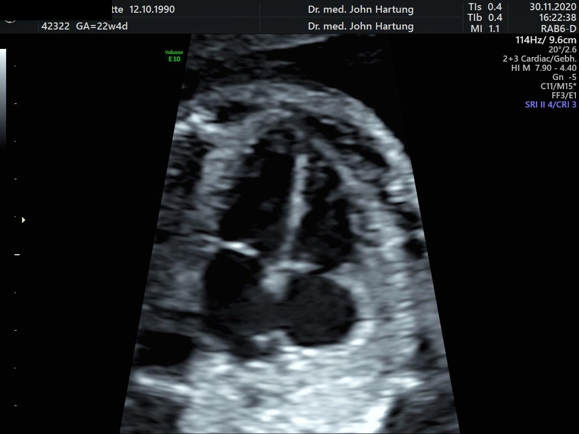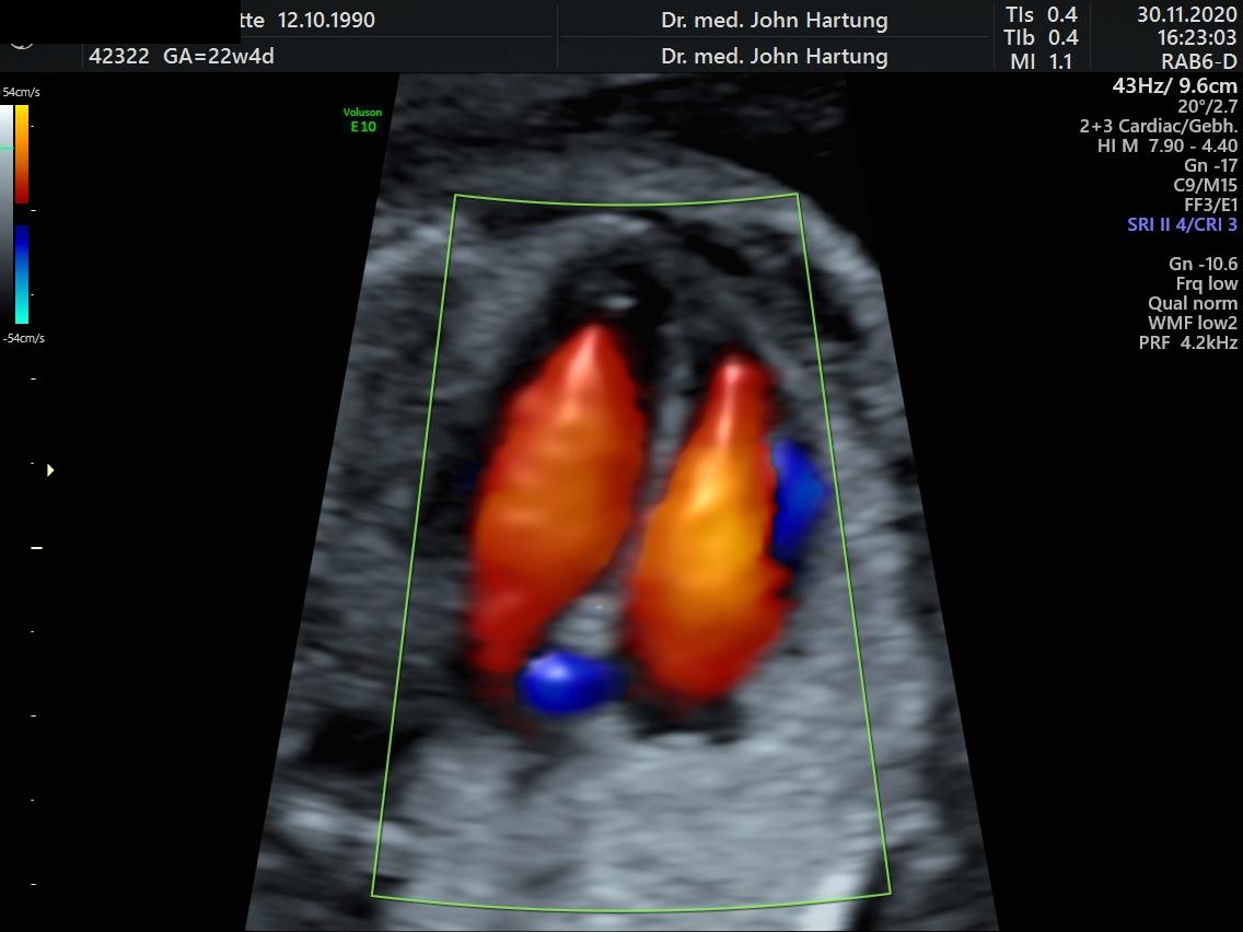


Fetal echocardiography
Fetal echocardiography
Congenital heart defects are among the most common malformations. They occur in 8 out of 1000 newborns. Most heart defects are serious in themselves, but they can also indicate other serious illnesses in the foetus. They therefore usually require specialised care of the newborn by paediatric cardiologists immediately after birth.
Foetal echocardiography is an ultrasound scan that examines the appearance, blood flow, and rate of the heart and nearby blood vessels. It can be performed using high-resolution equipment from the 13th week of pregnancy. However, the optimal time for the examination has been found to be between the 22nd and 24th week of pregnancy.
In more than 20% of heart defects, there are also chromosomal abnormalities. It may therefore be advisable to follow up unusual findings with a genetic examination in the form of an invasive procedure. Consultations with paediatric cardiologists and cardiac surgeons can be initiated before birth to ensure optimal care after birth.
Foetal echocardiography is a demanding examination and requires special equipment and training. As four-fifths of all heart defects in children are detected without any particular risks, this examination is actually always recommended, but it is strongly advised in the following cases:
- Diabetes or other metabolic diseases
- Taking certain medications in early pregnancy
- Congenital heart defects in the family
- monochorial twin pregnancies
- Suspicion of a heart defect or cardiac arrhythmia in the foetus
- Detection of other malformations of the foetus or increased nuchal translucency in the 11th-14th week of pregnancy
- Other risk situation