Fetale Echokardiographie

Fetale Echokardiographie

Zu den häufigsten Fehlbildungen gehören angeborene Herzfehler. Sie treten bei 8 von 1000 Neugeborenen auf. Herzfehler sind für sich schon größtenteils gravierend, sie können aber auch auf andere ernsthafte Erkrankungen des Feten hinweisen. Deshalb erfordern sie meist eine spezialisierte Betreuung des Neugeborenen durch Kinderkardiologen unmittelbar nach der Geburt.

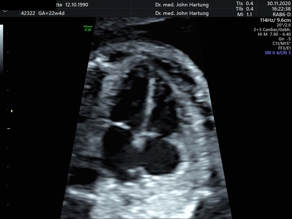
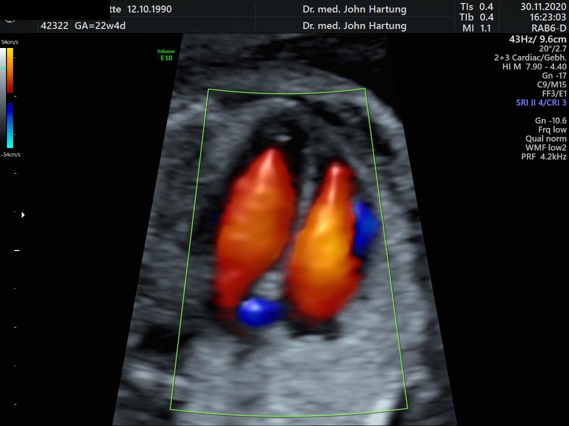
Die Fetale Echokardiographie ist eine Ultraschalluntersuchung, bei der das Aussehen, die Durchblutung und die Frequenz des Herzens und der herznahen Blutgefäße untersucht werden. Sie kann mit hoch auflösenden Geräten bereits ab der 13. Schwangerschaftswoche durchgeführt werden. Als günstigster Untersuchungszeitraum hat sich allerdings die 20. bis 22. Schwangerschaftswoche herausgestellt.

Bei mehr als 20% der Herzfehler bestehen auch chromosomale Auffälligkeiten. Deshalb kann es sinnvoll sein, bei ungewöhnlichen Befunden eine genetische Untersuchung in Form eines invasiven Eingriffs anzuschließen. Bereits vor der Geburt können Gespräche mit Kinderkardiologen und Herzchirurgen aufgenommen werden, um die optimale Betreuung nach der Geburt zu gewährleisten.

Die fetale Echokardiographie ist eine anspruchsvolle Untersuchung und erfordert eine spezielle Geräteausstattung und Ausbildung. Da vier Fünftel aller Herzfehler bei Kindern ohne besondere Risiken entdeckt werden, ist diese Untersuchung eigentlich immer empfehlenswert, in den folgenden Fällen ist sie allerdings dringend angeraten:

  • Diabetes oder anderen Stoffwechselerkrankungen
  • Einnahme bestimmter Medikamente in der Frühschwangerschaft
  • angeborenen Herzfehlern in der Familie
  • monochorialen Zwillingsschwangerschaften
  • Verdacht auf einen Herzfehler oder eine Herzrhythmusstörung des Feten
  • Nachweis anderer Fehlbildungen des Feten oder einer erhöhten Nackentransparenz in der 11.-14. SSW
  • anderweitige Risikosituation

Publikationen

Bamberg C, Hocher B, Slowinksi T, Halle H, Hartung J Pregnancy on intesified dialysis: fetal surveillance and outcome. Fetal Diagn Ther 2006   


Hartung J, Kalache KD, Heyna C,Heling K-S, Kuhlig M, Wauer R, Bollmann R, Chaoui R. Outcome of 60 neonates who had ARED flow prenatally

compared to a amtched control group of appropriate-for-gestational age preterm neonates. Ultrasound Obstet Gynecol 2005; 25:


Hartung J, Kalache K, Chaoui R. Der 3D- Power- Doppler- Ultraschall in der fetalen Diagnostik. Ultraschall in Med. (European J Ultrasound) 2004; 25: 195 – 199


Hartung J, Meckies J. Management of a case of uterine scar pregnancy treated by potassium injection. Ultrasound Obstet Gynecol 2003; 21 (1): 94 – 95 (LETTER)


Hartung J, Heling K-S, Rake A, Zimmer C, Chaoui R. Aneurysm of the vein of Galen detected in a 22 weeks fetus by signs of volume overload. Prenatal Diagn 2003; 23: 901 – 903


Urban M and Hartung J. Ultrasonographic and clinical appearance of a 22-week-old fetus with Brachmann-de-Lange Syndrome. Am J Med Gen 2001; 102 (1): 73-75


Chaoui R, Kalache K, Hartung J. Application of three dimensional power Doppler ultrasound in prenatal diagnosis. Ultrasound Obstet Gynecol 2001; 17: 22-29


Hartung J, Chaoui R, Bollmann. Non-immune hydrops secondary to fetomaternal hemorrage treated by serial fetal intravscular transfusions. Obstet Gynecol 2000; 96 (5): 844


Hartung J, Chaoui R, Kalache K, Tennstedt C, Bollmann R. Prenatal Diagosis of intrahepatic communications of the umbilical vein with atypical arteries (av-fistulae)
in two cases of trisomy 21 using color Doppler ultrasound. Ultrasound Obstet Gynecol 2000; 16 (3): 271 – 274


Hartung J, Chaoui R, Bollmann R. Amniotic fluid pressure in both cavities of twin – twin – transfusion syndrome. Fetal Diagn Ther 2000; 15 (2): 79-82


Hartung J, Enders G, Chaoui R, Arents, Tennstedt C, Bollmann R. Prenatal Diagnosis of congenital varicella syndrome and detection of the varicella virus in the fetus.
Prenatal Diagn 1999; 19 (2): 163 – 166


Hartung J, Chaoui R. Ultraschall in der Schwangerschaft: ein Beitrag zu einer sanften und sicheren Geburt. Der Gynäkologe (1999) 1: 60 – 65


Hartung J, Chaoui R, Wauer R, Bollmann R. Fetal hepatosplenomegaly: an isolated sign of trisomy 21 in a case of myeloprolipherative disorder.
Ultrasound Obstet Gynecol 1998; 11 (6): 453 – 455


Heling KS, Tennstedt C, Chaoui R, Kalache KD, Hartung J, Bollmann R. Reliability of prenatal sonographic lung biometry in the diagnosis of pulmonary hypoplasia.
Prenat Diagn 2001 21(8):649-57.


Heling K-S, Chaoui R, Hartung J, Kirchmair F, Bollmann R. Prenatal diagnosis of congenital neuroblastom. Fetal Diagn Ther 1999; 1: 47 – 52


Kalache KD, Chaoui R, Hartung J, Wernecke KD, Bollmann. Doppler assessment of tracheal fluid flow during fetal breathing movements in case of
congenital diaphragmatic hernia. Ultrasound Obstet Gynecol 1998; 12: 27 – 32


Chaoui R, Taddei F, Bast C, Lenz F, Kalache KD, Hartung J, Bollmann R. Sonographische Untersuchung des fetalen Lungenkreislaufs.
Der Gynäkologe 1997; 30: 230-239


Lun A, Lenz F, Priem F, Brux B, Gross J, Bollamnn R, Hartung J, Bartho S, Kirchmeier F, Reisinger I. Biochemical diagnosis in prenatal uropathy.
Clin Biochem 1994; 27 (4): 283 287


Lenz F, Machlitt A, Hartung J, Bollmann R, Chaoui R. Fetal pulmonary venous flow pattern is determined by left atrial pressure: a report of two cases of left heart hypoplasia,
one with patent and the other with closed interatrial communication. Ultrasound Obstet Gynecol 2002; 19 (4): 392 – 395


Hartung J, Tetzner V. Detection of muscular septal defects using Power – Doppler Ultrasound in mid – trimester echocardiography and description of natural history.
Ultrasound Obstet Gynecol 2004; 24: 218


Hartung J, Heyna C, Kalache KD, Heling K-S, Kuhlig M, Wauer R, Chaoui R, Bollmann R. Verlauf und Prognose von 60 Feten mit ARED –
Flow zwischen 24/0 und 34/0 SSW im Vergleich zu einer matched control Gruppe Frühgeborener. Ultraschall in der Medizin (European J Ultrasound) 2004; 562 – 563


Hartung J, Kalache KD, Heling K-S, Heyna C, Wauer R, Bollmann R, Chaoui R. Course and outcome of 60 fetuses with ARED-Flow between
24 and 34 weeks’ gestation compared to a matched control group. Ultrasound Obstet Gynecol 2003, Volume 22, Issue S1,32


Hartung J, Chaoui, R, Lenz, F, Kalache, K, Bollmann, R. Malformations of the intraabdominal umbilical vein: prenatal detection using color
Doppler and color power (1999) Archives of Perinatal Medicine Vol. 5, Suppl. 1, 11 – 12


Hartung J, Heling K-S, Rake A, Zimmer C, Chaoui R. Vein of Galen aneurysm detected at 22 weeks by cardiac signs of volume overload.
Ultrasound Obstet Gynecol 2002; 20:50


Hartung J, Chaoui, R, Kalache, K, Heling, K-S, Bollmann, R Abnormalities of the fetal umbilical vein: prenatal Diagnosis and fetal outcome (1998)
Ultrasound Obstet Gynecol Vol. 12, Suppl. 1, 52


Hartung J, Kalache, K, Chaoui, R, Bollmann, R. Amniotic fluid pressure during amniograinage in twin – to – twin transfusion syndrome and polyhydramnios (1998)
Ultrasound in Obstet Gynecol Vol. 12, Suppl. 1, 147


Hartung J, Chaoui, R, Bollmann, R.Is amniotic fluid pressure useful in predictig outcome in twin – to – twin syndrome ?
(1996) Ultrasound Obstet Gynecol (1996) Vol. 8, Supl. 1, 141


Hartung J, Chaoui, R, Bollmann, R.Hepatosplenomegaly in the fetus – a further marker of Trisomie 21 ? (1996) Ultrasound Obstet Gynecol Vol. 8, Supl. 1, 142


Chaoui, R, Kalache K. Heling K.-S., Hartung J, Bollmann R. Three – dimensional color power angiography in the assessment of fetal cardiovascular anatomy
(1999) Archives of Perinatal Medicine Vol. 5, Suppl. 1, 7


Chaoui R, Kalache K. Heling K.-S., Hartung J, Bollmann R. Three – dimensional color power angiography in the assessment of fetal cardiovascular anatomy
(1999) Archives of Perinatal Medicine Vol. 5, Suppl. 1, 7


Heling K.-S., Chaoui R. Hartung J, Kalache K, Bollmann R Perfusion through the foramen ovale in normal and in pregnancies complicated by IUGR and heart defects
(1999) Archives of Perinatal Medicine Vol. 5, Suppl. 1, 13


Lenz F, Chaoui R, Machlitt A, Hartung J, Heling K.S., Bollmann R Pulmonary venous flow in fetal cardiac anomalies (1999) Archives of Perinatal Medicine Vol. 5, Suppl. 1, 2


Heling, K-S, Chaoui, R, Hartung J, Kalache, K, Bollmann, R Hyperechogen Lungenfehlbildungen des Feten: Pränatale Diagnose und outcome bei 29 Feten (1999).
Z. Geburtsh. Neonatol. Vol. 203, 86


Heling, K-S, Hartung J, Chaoui, R, Kirchmeir, F, Bollmann, R Pränatale Diagnostik des fetalen Ileus (1999). Z. Geburtsh. Neonatol. Vol. 203, 87


Heling, K-S, Chaoui, R, Hartung J, Kalache, K, Bollmann, R. Z. Perfusion des Foramen ovale in der Schwangerschaft (1999) Z. Geburtsh. Neonatol. Vol. 203, 87


Lenz, F, Chaoui, R, Machlitt, A, Hartung J, Bollmann, R. Dopplersonographie der Pulmonalvenen bei fetalen Herzfehlern (1999) Z. Geburth. Neonatol. Vol. 203, 88


Kalache, K, Chaoui, R, Heling, K-S, Hartung J, Bollmann, R 3-D color Doppler in obstetrics: a useful alternativ to 3-D color power angio
(1998) Ultrasound Obstet Gynecol Vol. 12, Supl. 1, 56


Heling, K-S, Hartung J, Chaoui, R, Kalache, K, Bollmann, R Natural history of 23 fetuses with hyperechogenic lung lesions (1998). Ultrasound Obstet Gynecol Vol. 12, Supl. 1, 11


Heling, K-S, Chaoui, R, Hartung J, Kalache, K, Bollmann, R Fetal ovarian cyst – prenatal detection and postnatal outcome (1998) Ultrasound Obstet Gynecol Vol. 12, Supl. 1, 118


Heling, K-S, Chaoui, R, Hartung J, Kalache, K, Bollmann, R Biometry and Doppler values of the foramen ovale in the second half of pregncy
(1998) Ultrasound Obstet Gynecol Vol. 12, Supl. 1, 134


Chaoui, R, Kalache, K, Hartung J, Heling, K-S, Bollmann, R 3- D color Doppler angio (3-D CPA) of the umbilical vessels in normal and abnormal pregnancies
(1998) Ultrasound Obstet Gynecol Vol. 12, Supl. 1, 150


Hartung J, Tennstedt, C, Weidemeier, A, Bollmann, R. Oligohydramnios: sonographical differential diagnosis and prognosis
(1994), R. Pediatrics and Related Topics, 33 (4), 342 – 343