First trimester screening

First trimester screening

First of all: The vast majority of babies are healthy! Nevertheless, all women - regardless of their age - run a small risk of their baby being affected by a mental or physical illness.


The most common cause of mental disability is Down syndrome, a chromosomal disorder also known as trisomy 21. This disorder is not inherited, but occurs by chance. The risk of having a baby affected by Down syndrome increases with the age of the mother.


The only way to rule out a chromosomal disorder such as Down's syndrome with certainty is an invasive test, but this is not entirely harmless. You can find out more about these tests a few clicks further on.


However, many pregnant women do not want to make the decision for or against such invasive diagnostics solely on the basis of their age, but make it dependent on their individual risk.


First trimester screening is used to calculate the individual risk of Down's syndrome, to recognise serious malformations and pregnancy risks, and thus provides a basis for counselling for the entire course of the pregnancy.


Intensive research in recent years has shown that the majority of unborn babies affected by a chromosomal disorder show typical abnormalities in the early stages of pregnancy - either on ultrasound examination or in a maternal blood sample. 


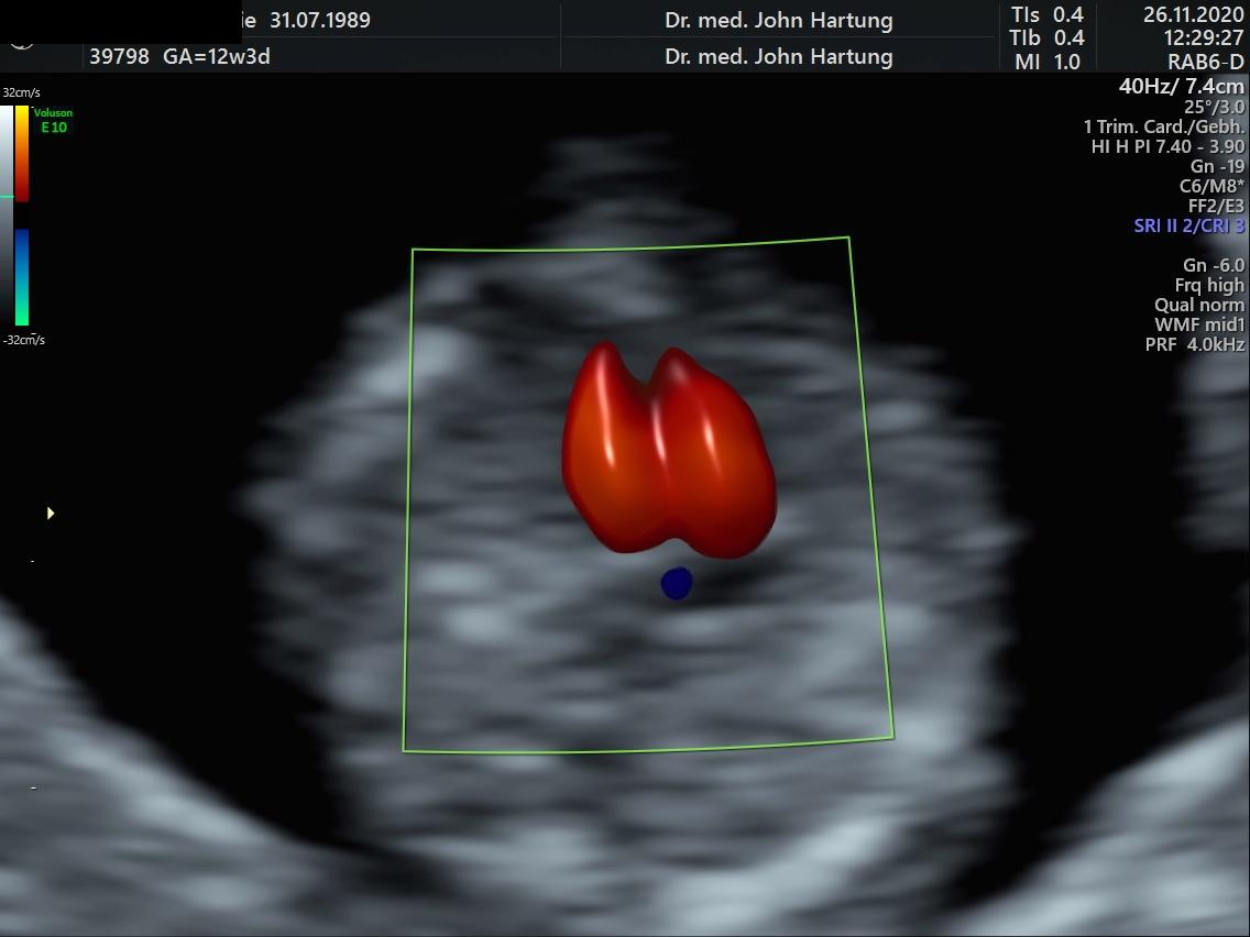
ULTRASOUND EXAMINATION

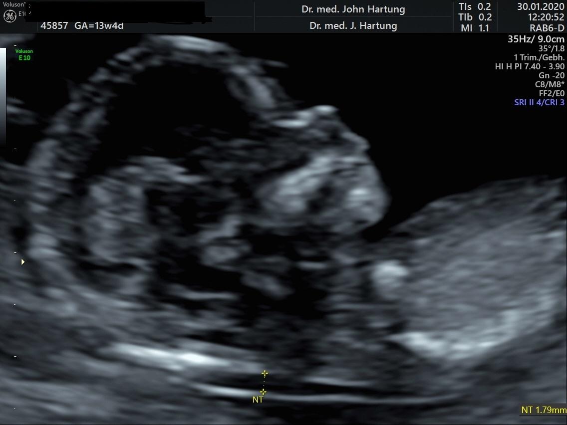
Between the 12th and 14th week of pregnancy, a small accumulation of fluid can be detected under the skin of the neck of every unborn child. The precise measurement of this fluid accumulation is called nuchal translucency measurement. An increased nuchal translucency increases the risk that the unborn child could be affected by a chromosomal disorder. In addition to nuchal translucency, other ultrasound findings can change the risk value of a chromosomal disorder.


To avoid any misunderstandings: The examination findings are only used to determine the risk and do not in themselves have any disease value, i.e. unborn babies with increased nuchal translucency and therefore an increased risk can of course also be completely healthy!

Blood test

Two values can be analysed from the mother's blood: the pregnancy hormone ß HCG and the protein PAPP-A produced by the placenta. The concentrations of these two substances - in addition to the ultrasound examination - determine the risk of a chromosomal disorder in the unborn child.


The blood test can also take place after the ultrasound scan, but the optimum time for the blood sample to be taken is earlier (10th to 11th week of pregnancy). In our practice, we calculate the risks together with the patients, questions and concerns regarding the examination can then be discussed.


Under optimal conditions, the combination of the three test components (maternal age, ultrasound examination, blood test) can detect over 90% of unborn babies with Down's syndrome, with the ultrasound examination alone over 80%.

Most examinations - even in older expectant mothers - reveal no abnormalities or low risks, which can help to reduce anxiety and ensure a carefree pregnancy. And that is worth a lot. 

To the forms
You are welcome to read through and complete the form on the subject in advance. 

Publications

Bamberg C, Hocher B, Slowinksi T, Halle H, Hartung J Pregnancy on intesified dialysis: fetal surveillance and outcome. Fetal Diagn Ther 2006   


Hartung JKalache KD, Heyna C,Heling K-S, Kuhlig M, Wauer R, Bollmann R, Chaoui R. Outcome of 60 neonates who had ARED flow prenatally

compared to an amtched control group of appropriate-for-gestational age preterm neonates. Ultrasound Obstet Gynecol 2005; 25:


Hartung JKalache K, Chaoui R. The 3D power Doppler ultrasound in foetal diagnostics. Ultrasound in Medicine (European J Ultrasound) 2004; 25: 195 - 199


Hartung JMeckies J. Management of a case of uterine scar pregnancy treated by potassium injection. Ultrasound Obstet Gynecol 2003; 21 (1): 94 - 95 (LETTER)


Hartung JHeling K-S, Rake A, Zimmer C, Chaoui R. Aneurysm of the vein of Galen detected in a 22 weeks foetus by signs of volume overload. Prenatal Diagn 2003; 23: 901 - 903


Urban M and Hartung J. Ultrasonographic and clinical appearance of a 22-week-old fetus with Brachmann-de Lange syndrome. Am J Med Gen 2001; 102 (1): 73-75


Chaoui R, Kalache K, Hartung J. Application of three dimensional power Doppler ultrasound in prenatal diagnosis. Ultrasound Obstet Gynecol 2001; 17: 22-29


Hartung JChaoui R, Bollmann. Non-immune hydrops secondary to fetomaternal hemorrage treated by serial fetal intravscular transfusions. Obstet Gynecol 2000; 96 (5): 844


Hartung JChaoui R, Kalache K, Tennstedt C, Bollmann R. Prenatal diagnosis of intrahepatic communications of the umbilical vein with atypical arteries (av-fistulae)
in two cases of trisomy 21 using colour Doppler ultrasound. Ultrasound Obstet Gynecol 2000; 16 (3): 271 - 274


Hartung JChaoui R, Bollmann R. Amniotic fluid pressure in both cavities of twin - twin - transfusion syndrome. Fetal Diagn Ther 2000; 15 (2): 79-82


Hartung JEnders G, Chaoui R, Arents, Tennstedt C, Bollmann R. Prenatal Diagnosis of congenital varicella syndrome and detection of the varicella virus in the fetus.
Prenatal Diagn 1999; 19 (2): 163 - 166


Hartung JChaoui R. Ultrasound in pregnancy: a contribution to a gentle and safe birth. The Gynaecologist (1999) 1: 60 - 65


Hartung JChaoui R, Wauer R, Bollmann R. Fetal hepatosplenomegaly: an isolated sign of trisomy 21 in a case of myeloprolipherative disorder.
Ultrasound Obstet Gynecol 1998; 11 (6): 453 - 455


Heling KS, Tennstedt C, Chaoui R, Kalache KD, Hartung JBollmann R. Reliability of prenatal sonographic lung biometry in the diagnosis of pulmonary hypoplasia.
Prenat Diagn 2001 21(8):649-57.


Heling K-S, Chaoui R, Hartung JKirchmair F, Bollmann R. Prenatal diagnosis of congenital neuroblastoma. Fetal Diagn Ther 1999; 1: 47 - 52


Kalache KD, Chaoui R, Hartung JWernecke KD, Bollmann. Doppler assessment of tracheal fluid flow during fetal breathing movements in case of
congenital diaphragmatic hernia. Ultrasound Obstet Gynecol 1998; 12: 27 - 32


Chaoui R, Taddei F, Bast C, Lenz F, Kalache KD, Hartung JBollmann R. Sonographic examination of the foetal pulmonary circulation.
The Gynaecologist 1997; 30: 230-239


Lun A, Lenz F, Priem F, Brux B, Gross J, Bollamnn R, Hartung JBartho S, Kirchmeier F, Reisinger I. Biochemical diagnosis in prenatal uropathy.
Clin Biochem 1994; 27 (4): 283 287


Lenz F, Machlitt A, Hartung JBollmann R, Chaoui R. Fetal pulmonary venous flow pattern is determined by left atrial pressure: a report of two cases of left heart hypoplasia,
one with patent and the other with closed interatrial communication. Ultrasound Obstet Gynecol 2002; 19 (4): 392 - 395


Hartung JTetzner V. Detection of muscular septal defects using Power - Doppler Ultrasound in mid - trimester echocardiography and description of natural history.
Ultrasound Obstet Gynecol 2004; 24: 218


Hartung J, Heyna C, Kalache KD, Heling K-S, Kuhlig M, Wauer R, Chaoui R, Bollmann R. Progression and prognosis of 60 foetuses with ARED -
Flow between 24/0 and 34/0 weeks' gestation compared to a matched control group of preterm infants. Ultrasound in Medicine (European J Ultrasound) 2004; 562 - 563


Hartung JKalache KD, Heling K-S, Heyna C, Wauer R, Bollmann R, Chaoui R. Course and outcome of 60 foetuses with ARED-Flow between
24 and 34 weeks' gestation compared to a matched control group. Ultrasound Obstet Gynecol 2003, Volume 22, Issue S1,32


Hartung JChaoui, R, Lenz, F, Kalache, K, Bollmann, R. Malformations of the intraabdominal umbilical vein: prenatal detection using colour
Doppler and colour power (1999) Archives of Perinatal Medicine Vol. 5, Suppl. 1, 11 - 12


Hartung JHeling K-S, Rake A, Zimmer C, Chaoui R. Vein of Galen aneurysm detected at 22 weeks by cardiac signs of volume overload.
Ultrasound Obstet Gynecol 2002; 20:50


Hartung JChaoui, R, Kalache, K, Heling, K-S, Bollmann, R Abnormalities of the fetal umbilical vein: prenatal diagnosis and fetal outcome (1998)
Ultrasound Obstet Gynecol Vol. 12, Suppl. 1, 52


Hartung J, Kalache, K, Chaoui, R, Bollmann, R. Amniotic fluid pressure during amniograinage in twin - to - twin transfusion syndrome and polyhydramnios (1998)
Ultrasound in Obstet Gynecol Vol. 12, Suppl. 1, 147


Hartung JChaoui, R, Bollmann, R.Is amniotic fluid pressure useful in predictig outcome in twin - to - twin syndrome ?
(1996) Ultrasound Obstet Gynecol (1996) Vol. 8, Supl. 1, 141


Hartung JChaoui, R, Bollmann, R.Hepatosplenomegaly in the fetus - a further marker of trisomy 21 ? (1996) Ultrasound Obstet Gynecol Vol. 8, Supl. 1, 142


Chaoui, R, Kalache K. Heling K.-S., Hartung J, Bollmann R. Three - dimensional colour power angiography in the assessment of fetal cardiovascular anatomy
(1999) Archives of Perinatal Medicine Vol. 5, Suppl. 1, 7


Chaoui R, Kalache K. Heling K.-S., Hartung J, Bollmann R. Three - dimensional colour power angiography in the assessment of fetal cardiovascular anatomy
(1999) Archives of Perinatal Medicine Vol. 5, Suppl. 1, 7


Heling K.-S., Chaoui R. Hartung J, Kalache K, Bollmann R Perfusion through the foramen ovale in normal and in pregnancies complicated by IUGR and heart defects
(1999) Archives of Perinatal Medicine Vol. 5, Suppl. 1, 13


Lenz F, Chaoui R, Machlitt A, Hartung J, Heling K.S., Bollmann R Pulmonary venous flow in fetal cardiac anomalies (1999) Archives of Perinatal Medicine Vol. 5, Suppl. 1, 2


Heling, K-S, Chaoui, R, Hartung J, Kalache, K, Bollmann, R Hyperechogenic lung malformations of the foetus: Prenatal diagnosis and outcome in 29 fetuses (1999).
Z. Obstetr. Neonatol. Vol. 203, 86


Heling, K-S, Hartung JChaoui, R, Kirchmeir, F, Bollmann, R Prenatal diagnosis of foetal ileus (1999). Z. Obstetr. Neonatol. Vol. 203, 87


Heling, K-S, Chaoui, R, Hartung JKalache, K, Bollmann, R. Z. Perfusion of the foramen ovale in pregnancy (1999) Z. Geburtsh. Neonatol. Vol. 203, 87


Lenz, F, Chaoui, R, Machlitt, A, Hartung JBollmann, R. Doppler sonography of the pulmonary veins in foetal heart defects (1999) Z. Geburth. Neonatol. Vol. 203, 88


Kalache, K, Chaoui, R, Heling, K-S, Hartung J, Bollmann, R 3-D colour Doppler in obstetrics: a useful alternative to 3-D colour power angio
(1998) Ultrasound Obstet Gynecol Vol. 12, Supl. 1, 56


Heling, K-S, Hartung JChaoui, R, Kalache, K, Bollmann, R Natural history of 23 foetuses with hyperechogenic lung lesions (1998). Ultrasound Obstet Gynecol Vol. 12, Supl. 1, 11


Heling, K-S, Chaoui, R, Hartung J, Kalache, K, Bollmann, R Fetal ovarian cyst - prenatal detection and postnatal outcome (1998) Ultrasound Obstet Gynecol Vol. 12, Supl. 1, 118


Heling, K-S, Chaoui, R, Hartung J, Kalache, K, Bollmann, R Biometry and Doppler values of the foramen ovale in the second half of pregnancy
(1998) Ultrasound Obstet Gynecol Vol. 12, Supl. 1, 134


Chaoui, R, Kalache, K, Hartung J, Heling, K-S, Bollmann, R 3- D colour Doppler angio (3-D CPA) of the umbilical vessels in normal and abnormal pregnancies
(1998) Ultrasound Obstet Gynecol Vol. 12, Supl. 1, 150


Hartung J, Tennstedt, C, Weidemeier, A, Bollmann, R. Oligohydramnios: sonographic differential diagnosis and prognosis
(1994), R. Pediatrics and Related Topics, 33 (4), 342 - 343